
Call For Appointment +91 8369683166


Protienuric Kidney Disease (Nephrotic & Nephritic Syndrome) & Renal Biopsy Procedure
- Home / Dr. Siddharth Lakhani
- Our Services
- Medical Management of Renal Transplant & it's Complications
- Medical Management of Chronic Kidney Disease & it's Complications
- Medical Management of Acute Kidney Injury
- Medical Management of Hemodialysis & Peritoneal Dialysis
- Kidney Stones & it's Medical Management & Prevention of Recurrence
- Hypertension: Primary, Secondary & Resistant Hypertension
- End Stage Renal Disease & its Management
- Diabetes, Hypertension, Pregnancy & Stone Related Kidney Disease
- Preventive Nephrology Services
- Protienuric Kidney Disease (Nephrotic & Nephritic Syndrome) & Renal Biopsy Procedure
- Glomerulonephritis & Management
- Electrolyte & Acid Base Imbalance
- Intervention Nephrology: Dialysis Catheter (Temporary & Tunneled Catheter)
- Fistula Salvage Procedure
Protienuric Kidney Disease (Nephrotic & Nephritic Syndrome) & Renal Biopsy Procedure
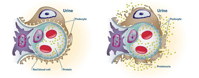
Proteinuric kidney diseases encompass conditions characterized by abnormal levels of protein in the urine. Nephrotic syndrome and nephritic syndrome are two distinct types of proteinuric kidney diseases, and a renal biopsy is often a crucial diagnostic tool for identifying the underlying cause and guiding treatment. Let's delve into each aspect
Nephrotic Syndrome
Characteristics: Nephrotic syndrome is characterized by significant proteinuria (loss of protein in the urine), hypoalbuminemia (low levels of albumin in the blood), edema (swelling), and hyperlipidemia (elevated levels of fats in the blood).
Causes: Common causes include minimal change disease, focal segmental glomerulosclerosis (FSGS), membranous nephropathy, and other glomerular disorders.
Renal Biopsy: A renal biopsy is often performed to determine the specific glomerular pathology and guide treatment decisions.
Renal Biopsy Procedure
Indications: Renal biopsy is typically indicated when proteinuria is persistent or severe, and the underlying cause needs to be identified for targeted treatment.
Procedure: A renal biopsy involves the removal of a small piece of kidney tissue for examination. This is usually done using a thin needle, guided by imaging techniques such as ultrasound or CT scan.
Complications: While renal biopsy is generally considered safe, there are potential complications, including bleeding and infection. Close monitoring is crucial after the procedure.
Pathological Examination: The biopsy sample is examined under a microscope to identify changes in the kidney tissue, helping in the diagnosis of the specific kidney disease.
Treatment
- Treatment of proteinuric kidney diseases depends on the underlying cause. It may involve medications to reduce inflammation, manage blood pressure, and address specific immune system abnormalities.
- In some cases, immunosuppressive therapies may be used to slow down or halt the progression of the disease.
Individualized management plans are crucial, and renal biopsy plays a central role in tailoring treatment to the specific pathology of the kidney disease. Close collaboration between nephrologists, pathologists, and other healthcare providers is essential for optimal patient care.